Skeletal Anatomy & Fractures
Skull, Le Fort, fracture types
When evaluating a trauma patient, specific mechanisms of injury are associated with predictable injury patterns. Always factor in the patient's age, use of restraints, presence of airbags, helmets, mechanism type, and estimated velocity when forming your clinical picture.
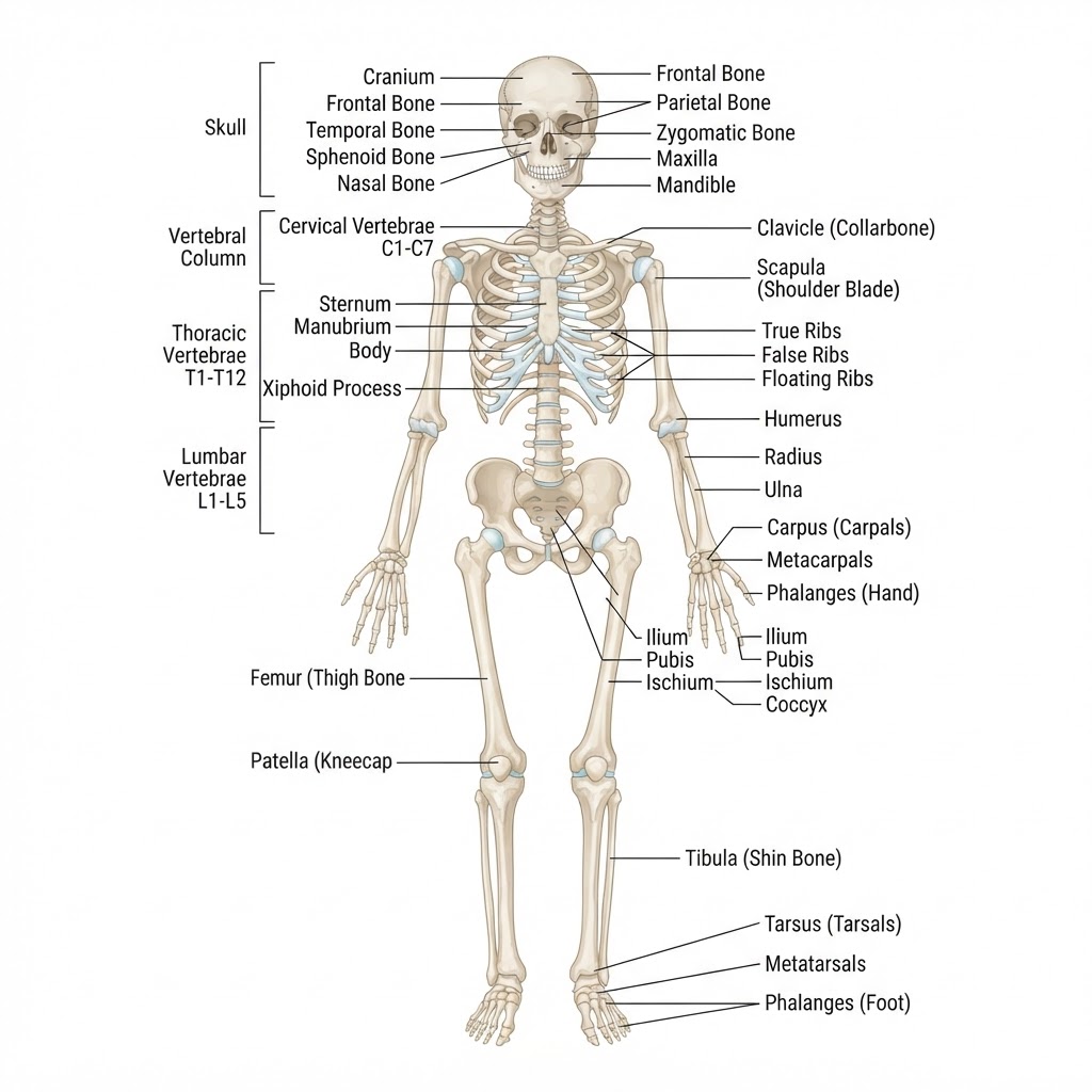
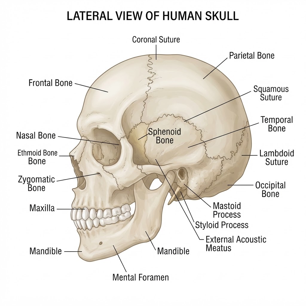
Lateral cranial and facial bone anatomy
Cranium
8 bones (frontal, parietal×2, temporal×2, occipital, sphenoid, ethmoid)
Facial Bones
14 bones including nasal, lacrimal, zygomatic, maxilla, mandible
Mandible
Only moveable facial bone; 1 bone
Vomer
Single midline nasal septum bone
Fracture description includes the bone, anatomical location, affected portion, fracture type, geometric orientation (transverse, oblique), and displacement subgroup (angulation, shortening, rotation).
Le Fort I
Horizontal maxillary fracture just above the teeth and palate, extending from the piriform fossa across the maxilla to the pterygoid fissure. May occur in isolation or alongside Le Fort II and III.
Le Fort II
Pyramidal fracture extending from the nasal dorsum across the medial orbital walls, through the maxilla below the zygomatic bone to the pterygomaxillary fissure.
Le Fort III
High-level craniofacial dissociation — fracture line extends from the nasal dorsum and cribriform plate along the medial and lateral orbital walls, detaching the zygomas from the cranial base.
- Battle's Sign — bruising behind the ear over the mastoid process; caused by fracture of the petrous portion of the temporal bone. Blood or CSF may drain from the ear (CSF otorrhea)
- Bulls-Eye Test — dab drainage from an orifice onto 4×4 gauze; a pinkish ring surrounded by clear fluid confirms CSF mixed with blood
- Raccoon Eyes — periorbital ecchymosis from anterior cranial fossa fracture; CSF may drain into sinuses; risk of olfactory and optic nerve damage
← swipe to navigate →